

Support Group for Pathologic Analysis
Head of the Group
Shinya Toyokuni (Nagoya University)
Support Activity
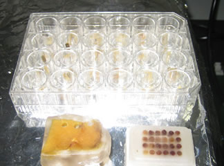
We provide cutting-edge pathologic analyses on rodents and other species. We consist of 7 registered pathologists, and can cover all sorts of pathologic alterations, including neoplasia, inflammatory conditions and neurodegenerative diseases. We can perform not only routine pathologic investigation (hematoxylin and eosin staining) but also special staining, immunohistochemistry, in situ hybridization and electronmicroscopic analysis. We can also conform to the analysis of iPS cell-derived animals.
Characteristics
Our major analyses focus on morphological alterations after drug (chemical) administration, and the phenotypic investigation on genetically engineered animals. Because 7 members cover the whole area of pathology, we are flexible enough to find the your person in charge, providing a high possibility of finding the phenotypes of genetically engineered animals. We include members who have abundant experience on iPS cells and genetically engineered animals with recently developed techniques. We can also provide the detailed analysis of fetus. We usually provide with preparation of pathologic specimens from tissues with detailed diagnosis, but only preparation of pathologic specimens or producing virtual slides is also available.
Examples
- Correct and detailed pathologic diagnosis for the macroscopic alterations in animals
- Cause of animal death
- Corresponding disease name as human counterparts
- Confirmation that there is no morphological alterations in the genetically engineered animals
- Cause of fetal death
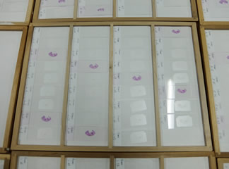
- Production of tissue microarray
- Reproducible immunohistochemistry
- Dissection of morphologically distinct cells
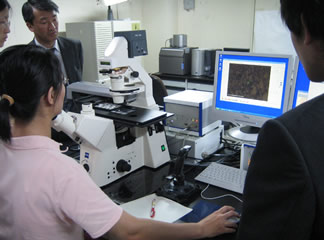
- Digitalization of pathologic information into virtual slides
- AI(artificial intelligence)-based morphological analysis of WSI(whole slide imaging) files
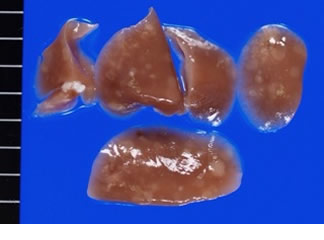 Starting from macroscopic findings |
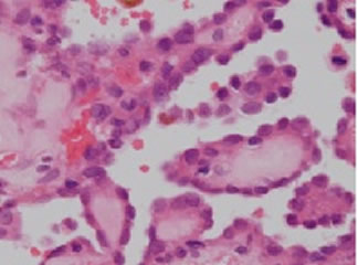 Histology with accurate diagnosis |
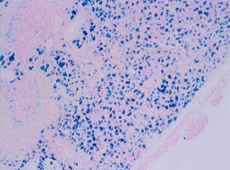 Important information with special staining |
 Analysis of embryo with serial sections |
 Production of tissue array from multiple samples |
 Laser microdissection |
Members for this support
Shinya Toyokuni (Nagoya University))
Mitsuru Futakuchi (Yamagata University)
Masaki Ueno (Kagawa University)
Tatsuhiko Miyazaki (Gifu University)
Hiroaki Kanda (Saitama Cancer Center)
Manabu Takamatsu (Cancer Institute)
Shugo Suzuki (Osaka Metropolitan University)
Application Form